Overview
At M.K Heart Clinic, our experts make use of the commonly used Intracoronary imaging techniques such as (OCT) Optimal Coherence Tomography and (IVUS) Intravascular Ultrasound in Chennai to visualize the inside of coronary arteries, and the blood vessels that supply the heart muscle with oxygen and nutrients.
- Intravascular Ultrasound is a medical imaging technique used to visualize the interior of blood vessels. It uses high-frequency sound waves to produce detailed images of the blood vessels, helping to detect blockages, plaque build-up, and other abnormalities. Intravascular Ultrasound in Chennai is performed during certain procedures, such as angioplasty and stenting, to guide treatment and improve outcomes.
- Optical Coherence Tomography is an imaging technique that uses light waves to create high-resolution, cross-sectional images of the coronary arteries. A small catheter with a light source and optical sensors is inserted into the coronary artery, and light is used to create images of the arterial walls and surrounding tissue. This makes it easier to see fine details such as the presence of plaque build-up or other abnormalities.
Cause
The primary cause of the use of intracoronary imaging techniques such as Intravascular Ultrasound and Optical Coherence Tomography in Chennai is to diagnose and treat coronary artery disease.
Coronary artery disease occurs when the coronary arteries become narrowed or blocked by plaque, a build-up of fat, cholesterol, and other substances in the blood. This can reduce or stop the flow of blood to the heart, leading to chest pain (angina), heart attack, or other complications.
Diagnosis
Intracoronary imaging techniques such as Intravascular Ultrasound and Optical Coherence Tomography in Chennai are used to diagnose and monitor various conditions affecting the coronary arteries, including coronary artery disease (CAD), stent deployment, arterial wall abnormalities, and vascular grafts.
The diagnostic procedures for Intravascular Ultrasound in Chennai are performed by
- Angioplasty.
- Stenting.
- Atherectomy.
- Drug-eluting stents.
Treatment at Intravascular Ultrasound in Chennai
The procedure for intracoronary imaging techniques such as Optical Coherence Tomography and Intravascular Ultrasound in Chennai typically involves the following steps:
- Before the Intravascular Ultrasound procedure, the patient is given a sedative, and a local anaesthetic is applied to the area where the catheter will be inserted.
- A long, thin catheter is inserted into an artery, usually in the groin, and threaded through the blood vessels to the heart. The catheter is equipped with an ultrasound transducer or optical fiber that is used to create images of the coronary arteries.
- Once the catheter is in place, the ultrasound transducer or optical fiber is activated and images are taken of the coronary arteries. The images can be displayed on a monitor in real-time, allowing the physician to assess the degree of blockage and guide interventional procedures.
- If needed, interventional procedures such as angioplasty, stenting, or atherectomy can be performed based on the images obtained from IVUS or OCT.
- After the procedure, the catheter is carefully removed and pressure is applied to the insertion site to prevent bleeding.
The entire procedure typically takes about 30-60 minutes and is performed in a cardiac catheterization laboratory or interventional radiology suite.
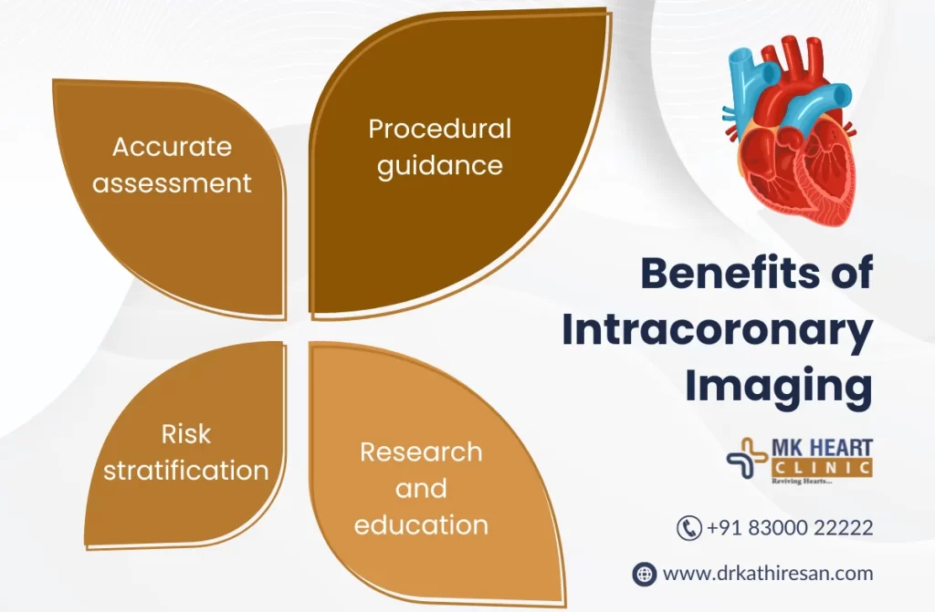
Benefits
The benefits of intracoronary imaging – Optical Coherence Tomography/ Intravascular Ultrasound in Chennai include:
- Improved Diagnosis.
- Guided Interventional Procedures.
- Reduced Invasiveness.
- Improved Outcomes.
Risks
Risks associated with Optical Coherence Tomography or Intravascular Ultrasound procedure include:
- Bleeding.
- Infection.
- Damage to Blood Vessels.

Facilities
- Cardiac Catheterization Laboratory or Interventional Radiology Suite: Intracoronary imaging techniques are typically performed in a cardiac catheterization laboratory or interventional radiology suite. These facilities are equipped with specialized imaging equipment, monitoring systems, and other equipment required to perform the procedure.
- Operating Room: In some cases, Optical Coherence Tomography or Intravascular Ultrasound in Chennai may be performed in an operating room under general anaesthesia.
Overall IVUS and OCT require specialized technology and facilities to perform, but the benefits in terms of improved diagnosis and treatment of heart disease make them an important tool for healthcare providers.
Our Doctors
Overall, Dr. M. Kathiresan plays a critical role in the process of Optical Coherence Tomography/ Intravascular Ultrasound in Chennai. This helps him to diagnose heart disease and recommend and guide effective treatment besides ensuring the best possible outcomes with a sophisticated treatment.